
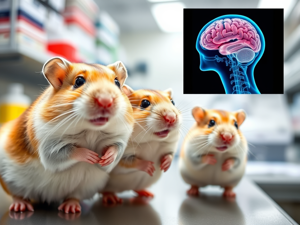
Cross-validation reveals fundamental contradictions between hamster model and human reality
Viral persistence claims directly contradicted by human autopsy evidence
The most striking contradiction comes from large-scale human autopsy studies that consistently fail to find SARS-CoV-2 in brain tissue. Columbia University’s comprehensive study of 41 COVID-19 patients found zero evidence of virus in brain cells despite using multiple detection methods including RNA in situ hybridization, viral protein antibodies, and RT-PCR across two dozen brain regions. Similarly, a New England Journal of Medicine study of 18 consecutive patients detected viral protein in exactly zero cases via immunohistochemistry, with only minimal RNA fragments found in 6 brain sections from 5 patients.
The Mayo Clinic’s autopsy series provides perhaps the most damning contradiction: among 72 patients tested, 98% showed negative viral detection, with the remaining 2% classified as indeterminate rather than positive. These findings directly contradict the Pasteur study’s claim of consistent viral detection at 80 days post-infection, with infectious virus isolated from 75-87.5% of hamster brains.
Multiple systematic reviews reinforce this pattern. A comprehensive 2021 analysis concluded that direct SARS-CoV-2 invasion of the central nervous system “is considered to be rare” based on accumulated autopsy evidence. The contrast is stark: while the Pasteur team reported successful viral isolation and cytopathic effects in cell culture from hamster brainstem tissue at 80 days, human studies using equally sensitive methods consistently report absent or minimal viral detection even at much earlier timepoints.
Animal model fundamentally fails to replicate human disease severity
A systematic review analyzing 27 COVID-19 animal model studies published in Critical Care revealed that no animal models, including hamsters, replicated severe COVID-19 features. The review found no models developed hypoxemic respiratory failure, multiple organ dysfunction, cytokine storm, or mortality – all hallmarks of severe human COVID-19. Instead, all animal models showed only mild disease with full recovery, suggesting what researchers termed a “wide gap between COVID-19 in humans and animal models.”
This translation failure extends specifically to brain pathology. While the Pasteur hamsters showed mild astrogliosis and behavioral changes, human COVID-19 brain pathology presents dramatically different patterns including widespread diffuse alveolar damage, severe encephalitis in some cases, and variable neuroinvasion patterns not seen in hamster models. A WHO expert working group concluded definitively that “no animal model tested thus far entirely reflects human COVID-19.”
The biological basis for these differences includes distinct ACE2 receptor expression patterns between species, with hamsters showing lower expression in key cell types compared to humans. Hamster immune responses are also “more limited and less characterized” than even mouse models, severely restricting mechanistic inference to human disease.
Alternative mechanisms explain neurological symptoms without viral persistence
Compelling evidence demonstrates that COVID-related brain changes occur through mechanisms entirely independent of direct viral infection. Stanford and UK Biobank studies found that pandemic stress alone caused measurable brain aging in non-infected individuals – with brains aging 5.5 months faster during the pandemic regardless of infection status. Adolescent brains showed cortical thinning equivalent to several years of aging even without COVID-19 infection.
The cytokine storm mechanism provides a particularly strong alternative explanation. Studies show COVID-19 patients exhibit the same metabolic brain disturbances as non-COVID hypoxic patients, but with cerebrospinal fluid remaining negative for SARS-CoV-2 RNA. This indicates peripheral inflammatory mechanisms rather than direct viral invasion. Autopsy studies consistently find widespread microglial activation but no viral RNA in brain tissue, supporting cytokine-mediated rather than viral-mediated damage.
Harvard MRI studies demonstrate that COVID patients show identical metabolic brain changes (NAA reduction, lactate elevation) to pure hypoxic brain injury, suggesting oxygen deprivation as the primary mechanism. The “silent hypoxia” phenomenon in COVID-19 – where patients experience oxygen desaturation without dyspnea – provides a clear pathway for brain damage without requiring viral neuroinvasion.
Dopamine pathway claims lack human validation
Columbia University neuropathologist James Goldman’s analysis of COVID autopsy brains provides definitive contradiction to the dopamine/Parkinson’s connection: “In the substantia nigra, the brain area that’s affected by Parkinson’s, there’s very little to see. There is no evidence of nerve cell degeneration.” He further emphasized finding “no pathology we can directly relate to the known pathology of Parkinson’s.”
The epidemiological evidence is equally contradictory. Despite billions of global COVID-19 infections, only approximately 20 cases of parkinsonism have been reported worldwide – a number the American Parkinson Disease Association calls “very small.” Laboratory studies seeking the pathological hallmark of Parkinson’s disease found no abnormally aggregated alpha-synuclein in cerebrospinal fluid from COVID-19 patients.
Recent research published in Cell demonstrates that long COVID brain fog is primarily linked to serotonin depletion, not dopamine dysfunction. This serotonin-based mechanism – involving gut inflammation, reduced tryptophan absorption, and vagus nerve dysfunction – was reversible with SSRIs in mouse models and has been confirmed by multiple independent studies.
Methodological issues compound translation problems
The Pasteur study’s own limitations further undermine translation to humans. The authors acknowledged they “do not show the direct causality between the persistent inflammation in the brain and the changed behaviors,” used behavioral tests “largely characterized in mice and rats” rather than validated for hamsters, and included “fewer animals than generally used for behavioral testing.”
The study’s controlled laboratory conditions using young, healthy hamsters starkly contrast with the elderly, comorbid human populations most affected by severe COVID-19. The 80-day endpoint, while impressive in hamster terms, may not reflect human disease trajectories given different lifespans and metabolic rates between species.
Human recovery evidence contradicts persistent damage narrative
Longitudinal human studies demonstrate recovery of COVID-related neurological symptoms, contradicting the persistent damage implied by the hamster model. Twelve-month follow-up studies show significant improvement in fatigue measures, with peripheral fatigue metrics improving from 37% to 55% over one year. Most post-COVID brain fog “clears up in the majority of people who have it” according to Yale Medicine, suggesting reversible rather than permanent changes.
Critical synthesis: The hamster model’s fundamental disconnect from human reality
The weight of contradictory evidence reveals that the Pasteur Institute’s hamster model, while potentially useful for basic viral biology research, fails to accurately represent human COVID-19 brain pathology. The claimed 80-day viral persistence in hamster brainstem stands in stark opposition to consistent negative findings in human autopsy studies using equally or more sensitive detection methods. The dopamine pathway effects observed in hamsters have no correlate in human neuropathological examinations.
Most critically, the alternative mechanisms identified – pandemic stress, cytokine storm, hypoxia, vascular damage, and autoimmunity – provide complete explanations for human neurological symptoms without requiring any viral persistence in brain tissue. The serotonin-based mechanisms for brain fog, supported by reversibility with treatment, further undermine the dopamine-centric narrative from the hamster model.
The Pasteur study exemplifies a broader pattern in COVID-19 research where animal model findings generate headlines but fail to translate to human clinical reality. For patients and clinicians, the evidence strongly supports focusing on treating the indirect effects of COVID-19 through anti-inflammatory, vascular, and supportive therapies rather than assuming persistent viral replication in the brain. The hamster model’s claims should be viewed as species-specific phenomena rather than predictive of human disease progression.
Major Articles and Studies Referenced in the Blog Post
Primary Study Discussed:
- Pasteur Institute Hamster Study (Nature Communications, July 2025)
- “Hamsters with long COVID present distinct transcriptomic profiles associated with neurodegenerative processes in brainstem”
- Authors: Coleon et al.
- Found viral persistence in hamster brainstem up to 80 days post-infection
Human Brain Autopsy Studies:
- Columbia University Study
- 41 COVID-19 patient brains examined
- Zero evidence of virus in brain cells
- Used RNA in situ hybridization, viral protein antibodies, and RT-PCR
- Mayo Clinic Autopsy Series
- 72 patients tested
- 98% showed negative viral detection
- Remaining 2% classified as indeterminate
- New England Journal of Medicine Study
- “Neuropathological Features of Covid-19”
- 18 consecutive patients
- Zero cases detected viral protein via immunohistochemistry
- Brain Autopsies of Critically Ill COVID-19 Patients (Acta Neuropathologica Communications)
- Demonstrated vascular injury and inflammation
- No viral RNA or protein detected in CSF or brains
Systematic Reviews and Meta-Analyses:
- Critical Care Systematic Review (2020)
- “Evidence of a wide gap between COVID-19 in humans and animal models”
- Analyzed 27 COVID-19 animal model studies
- Found no animal models replicated severe COVID-19 features
- 2021 Comprehensive Analysis
- Concluded direct SARS-CoV-2 CNS invasion is “considered to be rare”
Pandemic Brain Effects Studies:
- UK Biobank Study (Nature Communications, 2024)
- “Accelerated brain ageing during the COVID-19 pandemic”
- Found 5.5-month acceleration in brain aging regardless of infection status
- Stanford University Study (2022)
- “Pandemic stress physically aged teens’ brains”
- Showed cortical thinning in adolescents without COVID infection
Neurological Mechanism Studies:
- Columbia University Neuropathology
- Dr. James Goldman’s analysis
- Found no nerve cell degeneration in substantia nigra
- No Parkinson’s-like pathology
- Cell Journal Study
- Demonstrated long COVID brain fog linked to serotonin depletion
- Not dopamine dysfunction as suggested by hamster model
- Harvard MRI Studies
- Showed COVID patients have identical brain changes to hypoxic injury
- Suggested oxygen deprivation as primary mechanism
Clinical and Epidemiological Sources:
- American Parkinson Disease Association
- Reported only ~20 cases of COVID-related parkinsonism worldwide
- Called the number “very small” given billions of infections
- Yale Medicine
- Clinical guidance on long COVID brain fog
- Noted that symptoms clear up in majority of patients
- Michael J. Fox Foundation
- “News in Context: Does COVID cause Parkinson’s?”
- Analysis of COVID-Parkinson’s connection
Additional Key Studies:
- NIH Autopsy Study (Nature, December 2022)
- “SARS-CoV-2 infection and persistence in the human body and brain at autopsy”
- 44 patients studied
- Found virus throughout body but minimal brain involvement
- WHO Expert Working Group
- Concluded “no animal model tested thus far entirely reflects human COVID-19”
- Science for ME Discussion
- Critical analysis of the Pasteur hamster study limitations
- Noted methodological concerns
These articles represent a comprehensive cross-section of human autopsy studies, animal model critiques, alternative mechanism research, and clinical observations that contradict or contextualize the Pasteur Institute’s hamster findings.
